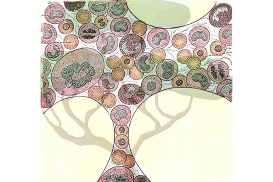
Blood cells make up the majority of cells in the human body. They perform critical functions and their dysfunction is implicated in many important human diseases, from anemias to blood cancers like leukemia. The many types of blood cells include red blood cells that carry oxygen, platelets that promote clotting, as well as the myriad types of immune cells that protect our bodies from threats such as viruses and bacteria.
What these diverse types of blood cells have in common is that they are all produced by hematopoietic stem cells (HSCs). HSCs must keep producing blood cells in large quantities throughout our entire lives in order to continually replenish our bodies’ supply. Researchers want to better understand HSCs and the dynamics of how they produce the many blood cell types, both in order to understand the fundamentals of human blood production and to understand how blood production changes during aging or in cases of disease.
Jonathan Weissman, an MIT professor of biology, member of the Whitehead Institute for Biomedical Research, and a Howard Hughes Medical Investigator; Vijay Sankaran, a Boston Children’s Hospital and Harvard Medical School associate professor who is also a Broad Institute of MIT and Harvard associate member and attending physician at the Dana Farber Cancer Institute; and Chen Weng, a postdoc in both of their labs, have developed a new method that provides a detailed look at the family trees of human blood cells and the characteristics of the individual cells, providing new insights into the differences between lineages of HSCs. The research, published in the journal Nature on Jan. 22, answers some long-standing questions about blood cell production and how it changes as we age. The work also demonstrates how this new technology can give researchers unprecedented access to any human cells’ histories and insight into how those histories have shaped their current states. This will render open to discovery many questions about our own biology that were previously unanswerable.
“We wanted to ask questions that the existing tools could not allow us to,” Weng says. “This is why we brought together Jonathan and Vijay’s different expertise to develop a new technology that allows us to ask those questions and more, so we can solve some of the important unknowns in blood production.”
How to trace the lineages of human cells
Weissman and others have previously developed methods to map the family trees of cells, a process called lineage tracing, but typically this has been done in animals or engineered cell lines. Weissman has used this approach to shed light on how cancers spread and on when and how they develop mutations that make them more aggressive and deadly. However, while these models can illuminate the general principles of processes such as blood production, they do not give researchers a full picture of what happens inside of a living human. They cannot capture the full diversity of human cells or the implications of that diversity on health and disease.
The only way to get a detailed picture of how blood cell lineages change through the generations and what the consequences of those changes are is to perform lineage tracing on cells from human samples. The challenge is that in the research models used in the previous lineage tracing studies, Weissman and colleagues edited the cells to add a trackable barcode, a string of DNA that changes a little with each cell division, so that researchers can map the changes to match cells to their closest relatives and reconstruct the family tree. Researchers cannot add a barcode to the cells in living humans, so they need to find a natural one: some string of DNA that already exists and changes frequently enough to allow this family tree reconstruction.
Looking for mutations across the whole genome is cost-prohibitive and destroys the material that researchers need to collect to learn about the cells’ states. A few years ago, Sankaran and colleagues realized that mitochondrial DNA could be a good candidate for the natural barcode. Mitochondria are in all of our cells, and they have their own genome, which is relatively small and prone to mutation. In that earlier research, Sankaran and colleagues identified mutations in mitochondrial DNA, but they could not find enough mutations to build a complete family tree: in each cell, they only detected an average of zero to one mutations.
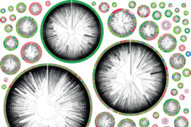
Now, in work led by Weng, the researchers have improved their detection of mitochondrial DNA mutations 10-fold, meaning that in each cell they find around 10 mutations — enough to serve as an identifying barcode. They achieved this through improvements in how they detect mitochondrial DNA mutations experimentally and how they verify that those mutations are genuine computationally. Their new and improved lineage tracing method is called ReDeeM, an acronym drawing from single-cell "regulatory multi-omics with deep mitochondrial mutation profiling." Using the method, they can recreate the family tree of thousands of blood cells from a human blood sample, as well as gather information about each individual cell’s state: its gene expression levels and differences in its epigenome, or the availability of regions of DNA to be expressed.
Combining cells’ family trees with each individual cell’s state is key for making sense of how cell lineages change over time and what the effects of those changes are. If a researcher pinpoints the place in the family tree where a blood cell lineage, for example, becomes biased toward producing a certain type of blood cell, they can then look at what changed in the cells’ state preceding that shift in order to figure out what genes and pathways drove that change in behavior. In other words, they can use the combination of data to understand not just that a change occurred, but what mechanisms contributed to that change.
“The goal is to relate the cell’s current state to its past history,” Weissman says. “Being able to do that in an unperturbed human sample lets us watch the dynamics of the blood production process and understand functional differences in hematopoietic stem cells in a way that has just not been possible before.”
Using this approach, the researchers made several interesting discoveries about blood production.
Blood cell lineage diversity shrinks with age
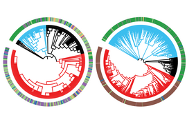
The researchers mapped the family trees of blood cells derived from each HSC. Each one of these lineages is called a clonal group. Researchers have had various hypotheses about how clonal groups work: Perhaps they are interchangeable, with each stem cell producing equivalent numbers and types of blood cells. Perhaps they are specialized, with one stem cell producing red blood cells, and another producing white blood cells. Perhaps they work in shifts, with some HSCs lying dormant while others produce blood cells. The researchers found that in healthy, young individuals, the answer is somewhere in the middle: Essentially every stem cell produced every type of blood cell, but certain lineages had biases toward producing one type of cell over another. The researchers took two samples from each test subject four months apart, and found that these differences between the lineages were stable over time.
Next, the researchers took blood samples from people of older age. They found that as humans age, some clonal groups begin to dominate and produce a significantly above-average percent of the total blood cells. When a clonal group outcompetes others like this, it is called expansion. Researchers knew that in certain diseases, a single clonal group containing a disease-related mutation could expand and become dominant. They didn’t know that clonal expansion was pervasive in aging even in seemingly healthy individuals, or that it was typical for multiple clonal groups to expand. This complicates the understanding of clonal expansion but sheds light on how blood production changes with age: The diversity of clonal groups decreases. The researchers are working on figuring out the mechanisms that enable certain clonal groups to expand over others. They are also interested in testing clonal groups for disease markers to understand which expansions are caused by or could contribute to disease.
ReDeeM enabled the researchers to make a variety of additional observations about blood production, many of which are consistent with previous research. This is what they hoped to see: the fact that the tool efficiently identified known patterns in blood production validates its efficacy. Now that the researchers know how well the method works, they can apply it to many different questions about the relationships between cells and what mechanisms drive changes in cell behavior. They are already using it to learn more about autoimmune disorders, blood cancers, and the origins of certain types of blood cells.
The researchers hope that others will use their method to ask questions about cell dynamics in many scenarios in health and disease. Sankaran, who is a practicing hematologist, also hopes that the method one day revolutionizes the patient data to which clinicians have access.
“In the not-too-distant future, you could look at a patient chart and see that this patient has an abnormally low number of HSCs, or an abnormally high number, and that would inform how you think about their disease risk,” Sankaran says. “ReDeeM provides a new lens through which to understand the clone dynamics of blood production, and how they might be altered in human health and diseases. Ultimately, we will be able to apply those lessons to patient care.”
