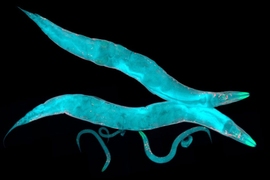
A new MIT study that focuses on a single cell in one of nature’s simplest nervous systems provides an in-depth illustration of how individual neurons can use multiple means to drive complex behaviors.
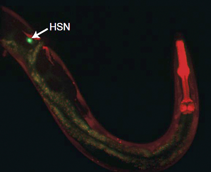
In the C. elegans worm, which only has 302 nerve cells, the neuron HSN releases several chemicals and makes multiple connections along its length to not only control the animal’s instantaneous egg laying and locomotion, but also to then slow the worm down for several minutes after the eggs are laid. To control that latter phase of the behavior, HSN transfers the neurotransmitter serotonin to a fellow neuron, which re-releases it to influence behavior minutes later.
“Our results reveal how a single neuron can influence a broad suite of behaviors over multiple timescales and show that neurons can ‘borrow’ serotonin from one another to control behavior,” the researchers report in Current Biology.
The study’s senior author is Steven Flavell, associate professor in The Picower Institute for Learning and Memory and the Department of Brain and Cognitive Sciences. Postdoc Yung-Chi Huang is the study’s first author.
A busy neuron
Heading into the study, HSN’s connections to other neurons had already been mapped (C. elegans is the only animal where the full connectome among neurons is known), and it had also been associated with egg-laying. Flavell’s lab, meanwhile, has observed that when the worms lay eggs, they speed across patches of food a bit like a farmer will drive a tractor to disperse seeds throughout fertile soil. Moreover, scientists had observed that when HSN is ablated, worms don’t engage in a characteristic feeding behavior of slowing down to dine on patches of food.
But how this single neuron had these seemingly paradoxical effects on behavior (egg laying, speeding up to do that, and slowing down after) remained a mystery. Flavell and Huang’s team employed a wide variety of techniques and experiments to discover how HSN does its many jobs.
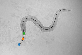
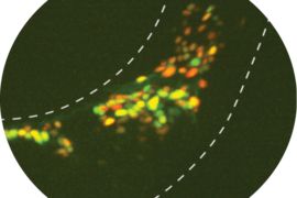
To establish that HSN indeed has a causal role in controlling these behaviors, they manipulated the neuron’s activity using optogenetics, a technique in which cells are genetically engineered to be controlled by flashes of light. In addition to confirming HSN’s key role in egg laying, these and other genetic manipulation experiments also confirmed that HSN causes the worm to speed up and then, after a bout of roaming and egg laying, to slow down for several minutes. The lab also tracked the electrical activity of HSN during these behaviors (by tracking the flow of calcium ions within the cell as animals freely moved) and saw that specific patterns of the cell’s activity were associated with egg laying and locomotion.
Many molecular means
Having established that HSN causes the three related behaviors, the lab then zeroed in on how it does so.
HSN is known to release a wide variety of neurotransmitter chemicals including serotonin, acetylcholine, and numerous peptides. Its release of serotonin and a neuropeptide called NLP-3 is known to drive egg-laying. To determine how the neuron drives speedy locomotion, the team systematically knocked out each HSN neurotransmitter and then stimulated HSN to see whether the worms could still speed up when HSN couldn’t produce one chemical or the other. The experiments revealed that HSN drives increased locomotion via the release of two neuropeptides, called FLP-2 and FLP-28.
Knocking out serotonin in HSN, meanwhile, disabled the worm’s slowing behavior. Further experiments showed how. Flavell’s team has previously studied the neuron NSM, showing that it uses serotonin when a worm is feeding to inhibit motor circuits and slow the worm down for the meal. In this study, the team showed that NSM’s action depended on a supply of serotonin coming from HSN. When HSN couldn’t produce serotonin, for instance, NSM couldn’t slow the worms down as well. The team further showed that NSM uses the serotonin transporter SERT (called MOD-5 in C. elegans) to take up HSN’s serotonin and re-release it. This showed that serotonergic neurons can pool and share serotonin with one another, with a direct impact on the animal’s behavior, Flavell says.
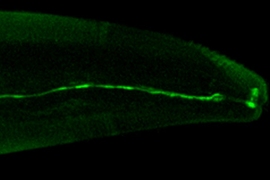
Turning to HSN’s anatomy, the team discerned that control of locomotion and control of egg-laying occurred along different points of HSN’s axon. HSN’s cell body is in the midbody of the animal. It forms synapses with the egg-laying circuit in the midbody and then its axon projects to the head to make synapses with other neurons. Cutting HSN’s axon between the midbody and head did not disrupt the animal’s egg-laying, but did prevent the coordination of egg-laying and locomotion, suggesting that HSN’s projection to the head coordinates HSN’s action on the egg-laying circuit with its action on the locomotion circuit.
In all, the study showed how HSN uses many parallel neurotransmitter outputs in different ways to control the animal’s behavior.
“Our results illustrate how cellular morphology, multiple transmitter systems, and non-canonical modes of transmission like neurotransmitter “borrowing” endow a single neuron with the ability to orchestrate multiple features of a behavioral program,” the authors wrote.
Meanwhile, the finding that neurons can take up and re-release serotonin produced by other neurons to control behavior reveals a novel feature of serotonin signaling that could have important medical implications, Flavell says. The molecule that takes up the serotonin, SERT/MOD-5, is the target of serotonin-specific reuptake inhibitors (SSRIs) like Prozac. This study raises the possibility that SSRIs may influence how neurons share serotonin with one another, which could be relevant for their mode of action in treating a wide variety of psychiatric disorders.
In addition to Huang and Flavell, the paper’s other authors are Jinyue Luo, Wenjia Huang, Casey Baker, Matthew Gomes, Bohan Meng, and Alexandra Byrne.
The National Institutes of Health, the National Science Foundation, the McKnight Foundation, The Alfred P. Sloan Foundation, The Picower Institute, and The JPB Foundation contributed funding for the study.
